
Understanding How X-Rays Reveal the Progression of Degenerative Changes in Your Body
由 Zestora 上 Feb 11, 2026

X-ray imaging is a vital tool used by healthcare providers to visualize the internal structure of the body, particularly to assess bone integrity and alignment. When patients undergo X-rays, they may encounter terminology such as 'degenerative changes.' This language reflects findings that can be associated with age, activity, and prior physical stress. Understanding how X-rays reveal these changes provides valuable context for patients as they navigate their health journeys. While X-rays can show structural details, it's important to know that they do not convey information about pain levels or overall functional ability. Therefore, recognizing the role of X-rays in monitoring progression and their limitations can empower patients to make informed decisions about their health management.

Key Takeaways
- X-rays are essential tools in medical imaging that help visualize changes within the body.
- Degenerative changes refer to the gradual deterioration of body structures, often associated with aging or injury.
- Understanding how X-rays work provides insight into their effectiveness in identifying these changes.
- Interpreting X-ray results helps healthcare professionals recognize the extent and nature of degenerative changes.
- Regular X-ray monitoring is crucial for tracking the progression of degenerative conditions and guiding treatment decisions.
Introduction to X-Rays and Their Importance in Medical Imaging
X-rays are a vital tool in medical imaging that provide healthcare professionals with important insights into the structure and alignment of bones and joints. When an X-ray is ordered by a licensed medical doctor, it is based on an individual’s specific history and physical examination, allowing for a tailored approach to the patient’s needs. The resulting images are interpreted by qualified medical professionals who analyze the findings within the broader context of the patient’s health and clinical background. As common terminology, phrases such as 'the X-ray shows progression of degenerative changes' may appear in reports, especially as individuals age or engage in long-term physical activities. However, it is essential to understand that X-rays depict structural aspects, not pain levels or functional abilities, meaning that the presence of such changes does not directly translate to any specific experience of discomfort or mobility. Ultimately, X-rays serve as one part of the overall medical assessment, guiding healthcare providers in their decisions regarding care.
What are Degenerative Changes? Understanding the Concepts
Degenerative changes refer to the natural wear and tear that can occur in joints and connective tissues over time, often visible in X-ray imaging. When an X-ray shows progression of degenerative changes, it indicates alterations in the structure and alignment of bones or joints, which may be influenced by factors such as age, long-term activity, or previous physical stress. While these findings are common, especially among older adults or individuals with a history of specific physical activities, it is important to remember that X-rays primarily illustrate the structural condition of bones rather than assess aspects like pain, function, or mobility. Thus, the presence of degenerative changes on an X-ray does not necessarily dictate an individual's level of comfort or ability to move. Understanding these concepts can help individuals process their imaging results within the broader context of their overall health and lifestyle.
'The doctors are the architects of human happiness; they build the foundations of health with the bricks of knowledge and skill, much like an X-ray reveals the unseen progression beneath the surface.' - Unknown
How X-Rays Work: Mechanisms Behind Imaging
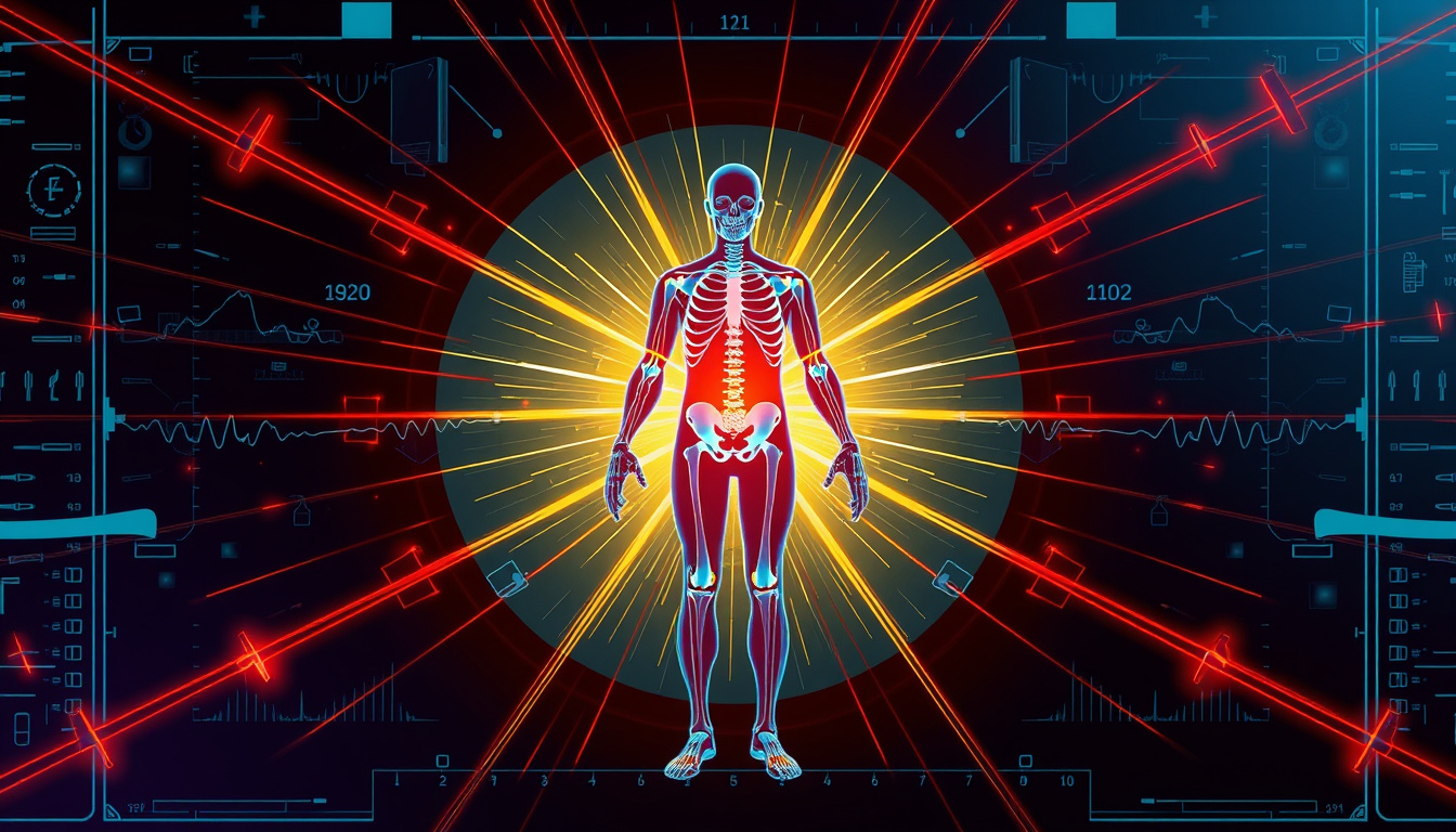
X-rays are a form of electromagnetic radiation that can penetrate soft tissue and are absorbed by denser materials like bones. When a patient undergoes an X-ray, the machine generates a controlled beam of radiation that passes through the body and captures images of the internal structures. This imaging technique is particularly useful in observing the skeletal system, as it clearly defines bone structure and alignment. When healthcare professionals refer to the 'progression of degenerative changes' in an X-ray report, they are noting observable structural alterations that can occur over time due to factors such as age, repetitive activity, or prior physical stress. It's important to recognize that these observations form only a part of a larger clinical picture, which also takes into account a person's history, physical examination, and overall function.
Interpreting X-Ray Results: What Degenerative Changes Look Like
X-ray imaging is an important tool used by licensed medical professionals to evaluate and assess the structural aspects of the body, particularly bones and joints. When X-ray reports mention terms such as 'degenerative changes,' it often refers to the natural aging process or the accumulation of wear and tear on joints that can occur over time. However, it is crucial to remember that while X-rays can provide valuable insights into bone structure and alignment, they do not offer a complete picture of a person’s overall health, comfort, or mobility. These imaging results are interpreted within the context of a patient's complete medical history and physical examination by healthcare providers. Thus, any interpretations regarding the 'progression of degenerative changes' should be viewed as part of a broader assessment rather than a definitive statement about an individual's current condition or functionality.

Monitoring Progression: How X-Rays Track Disease Development
X-ray imaging plays a crucial role in monitoring the progression of degenerative changes in various body structures, particularly in the context of conditions that may develop over time. When referred to as showing progression, it often refers to the comparative analysis of X-ray images taken at different points in time. Medical professionals evaluate these images, looking for changes in bone structure and alignment which may indicate how a person's condition is evolving. It is important to understand that while X-rays can provide valuable insights into structural changes, they do not directly correlate with symptoms such as pain or functionality. Therefore, a comprehensive assessment that includes clinical evaluation and patient reports is essential for a complete understanding of an individual's health status.
Conclusion: The Role of X-Rays in Managing Degenerative Conditions
In conclusion, X-ray imaging serves as an important tool in the assessment of degenerative conditions by providing insights into structural changes over time. When X-rays show progression of degenerative changes, this information can help licensed medical professionals evaluate an individual's condition and guide appropriate care. However, it is essential to remember that X-ray findings do not encapsulate the entire picture of a person's health or experience; factors such as pain levels, mobility, and overall function are critical to understanding an individual's quality of life. Consequently, many individuals consider additional supportive measures for joint and connective-tissue health, particularly as they age or engage in long-term physical activities. Integrating professional care with thoughtful nutritional support can play a role in promoting overall well-being.
- age related xray changes,
- alignment changes xray,
- alongside professional care,
- bone spurs xray,
- boswellia frankincense education,
- calcification xray,
- cats claw ingredient education,
- chronic xray changes,
- collagen type ii education,
- common xray terms,
- compression deformity xray,
- connective tissue support,
- decreased bone density xray,
- degenerative changes xray,
- disc space narrowing xray,
- enthesophyte xray,
- facet arthropathy xray,
- fda disclaimer supplements,
- foot ankle xray findings,
- ginger ingredient education,
- hand wrist xray findings,
- healed fracture xray,
- hip xray findings,
- hyaluronic acid education,
- imaging context education,
- imaging does not equal symptoms,
- incidental xray findings,
- ingredient education joints,
- interval change xray,
- joint space narrowing xray,
- joint structure education,
- knee xray findings,
- medical interpretation required,
- normal xray report,
- osteoarthritis xray terminology,
- osteopenia xray,
- osteophytes xray,
- scoliosis xray,
- shoulder xray findings,
- spinal spondylosis xray,
- spine xray findings,
- stable degenerative changes,
- subchondral sclerosis xray,
- supplement structure function,
- turmeric ingredient education,
- unremarkable xray,
- wear and tear xray,
- xray findings education,
- xray limitations,
- xray report explained


