
Understanding How Ultrasound Reveals Volar Plate Injuries: Diagnosis and Treatment Insights
by Zestora on Feb 12, 2026

Ultrasound imaging plays a vital role in diagnosing soft-tissue injuries, including those affecting the volar plate in the hand. Understanding the function of the volar plate and how ultrasound can be used to assess such injuries can provide important insights into hand function and overall recovery.
Key Takeaways
- The volar plate is a crucial ligament that supports hand function by stabilizing the finger joints.
- Volar plate injuries commonly occur due to trauma, sports injuries, or falls, impacting hand mobility.
- Ultrasound imaging provides a non-invasive method for accurately diagnosing volar plate injuries.
- Interpreting ultrasound results helps healthcare professionals identify the extent and specifics of the injury.
- Effective treatment and rehabilitation strategies are essential for recovery from volar plate injuries to restore function.
What is the Volar Plate and Its Role in Hand Function?
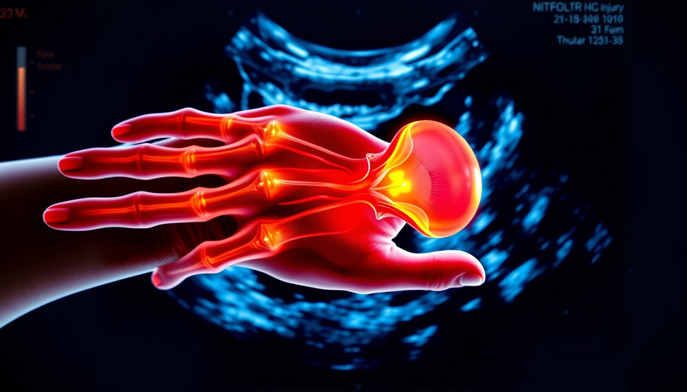
The volar plate is a crucial structure in the hand, often located at the base of the fingers. It is a thickened band of tissue that connects the bones at the finger joints, providing stability and support while allowing for smooth movement. When ultrasound shows a volar plate injury, this imaging technique captures any soft-tissue changes or abnormalities associated with the plate's condition, including structural appearance and the ability to move without restriction. The terminology around such injuries can help healthcare providers understand the extent of the involvement of the volar plate in hand function. However, it's noteworthy that descriptions from imaging alone do not convey how an individual may function or experience mobility in daily activities. As a result, some people consider both the results from imaging and the importance of supportive care, including nutritional elements, to maintain the health of their connective tissues and promote overall hand function.
Common Causes of Volar Plate Injuries
When an ultrasound shows a volar plate injury, it may indicate damage to this critical ligament located on the palm side of the finger. Common causes of such injuries often include traumatic events or repetitive stress, such as a fall onto an outstretched hand or engaging in activities that place strain on the finger joints. Athletes who participate in sports with high impacts or those that involve gripping, like basketball or gymnastics, might be more susceptible to these types of injuries. Understanding the potential causes and the language used in ultrasound reports can help individuals better comprehend their condition and the nature of the injury.
'The simple act of paying attention can take you a long way.' – Keanu Reeves
How Ultrasound Imaging Works in Diagnosing Volar Plate Injuries
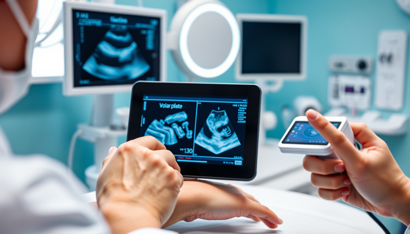
Ultrasound imaging is a valuable tool used by licensed medical professionals to visualize soft-tissue structures in the body, including conditions such as a volar plate injury. The ultrasound system employs sound waves to create real-time images of joints, tendons, ligaments, and surrounding tissues, allowing the clinician to assess movement and structure. It is important to know that while the ultrasound can show the presence of structural changes related to a volar plate injury, such descriptions do not reflect the patient's pain levels or functional capabilities. An ultrasound may reveal findings consistent with a volar plate injury despite the individual maintaining a reasonable level of function. The findings from an ultrasound, therefore, serve as part of a broader assessment, and any diagnosis or care plan should always be discussed with a qualified healthcare provider.
Interpreting Ultrasound Results: Identifying Specific Injuries
When ultrasound reports indicate findings such as a 'volar plate injury,' it reflects the ability of ultrasound to visualize soft-tissue structures and dynamics within the body. The volar plate is a ligamentous structure found on the palm side of the fingers that plays an important role in joint function. Ultrasound imaging focuses on the appearance and movement of this soft tissue, describing its structure or any potential deviations from the norm. However, it is essential to note that these findings do not directly correlate with pain levels or functional capabilities. Many individuals may exhibit structural changes without experiencing significant limitations in their daily activities. Therefore, discussions about ultrasound results should always take place with a qualified healthcare provider who can interpret these findings in the context of an individual’s overall health and functional goals.
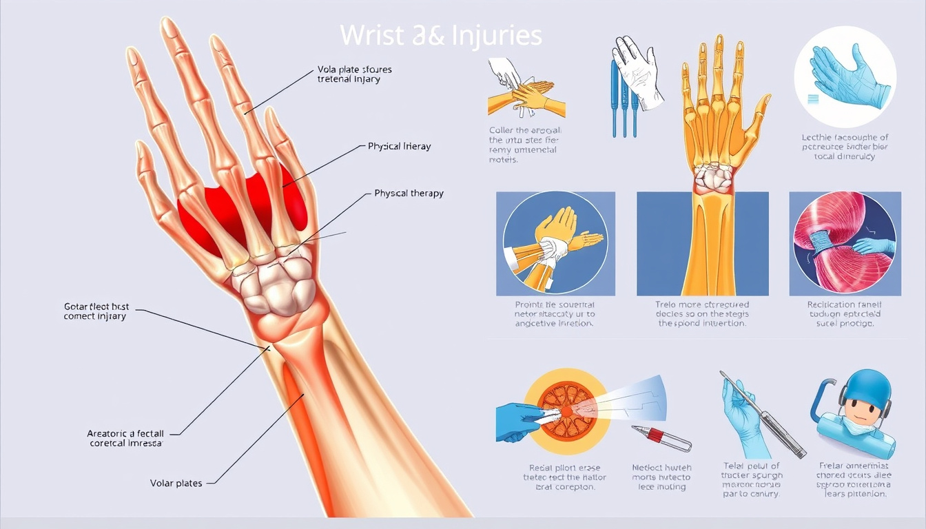
Treatment Options for Volar Plate Injuries: A Comprehensive Overview
Ultrasound shows volar plate injury, a common finding in imaging reports, particularly in cases involving hand injuries. The volar plate is a fibrocartilaginous structure that stabilizes the fingers and helps with their range of motion. When an ultrasound is conducted, it allows for a dynamic view of this soft-tissue structure as well as its movement, providing essential insight into the injury. Licensed medical professionals interpret these ultrasound results in conjunction with physical assessments to guide treatment decisions. It is important to understand that the findings captured in an ultrasound report focus on the appearance and potential movement of the volar plate, rather than direct measures of pain or functional capability. Although an ultrasound shows a volar plate injury, individuals can still maintain a relatively good function in their fingers, as structural findings do not always correlate directly with a person's lived experience or capabilities in daily activities. Consequently, some individuals may choose to focus on long-term support of the affected tissue, considering factors like gradual adaptation and consistency in their rehabilitation to reinforce normal structure and function of the joint.
Rehabilitation and Recovery Process After Volar Plate Injury
When ultrasound reports mention findings related to a volar plate injury, it typically indicates that the imaging has been ordered by a licensed medical professional to assess the condition of the soft tissues associated with the finger joint. Ultrasound is particularly effective at visualizing the appearance and movement of these types of structures, such as the volar plate itself, along with associated tendons and ligaments. It is important to note that the findings presented in ultrasound reports primarily focus on structural aspects or motion rather than providing information about pain, strength, or an individual’s ability to perform daily activities. Therefore, it is possible for ultrasound findings to reveal a volar plate injury in a person who still maintains a relatively good level of function, as the structure of soft tissues does not always correlate directly with a person’s lived experience of capability. Moreover, some individuals may choose to focus on long-term tissue support and maintenance strategies, recognizing that gradual adaptation and consistent care can help support normal tissue structure over time. This approach often includes nutritional support, where nutrients like Type II collagen, turmeric, ginger, and hyaluronic acid are commonly discussed for their potential roles in supporting normal joint and connective tissue structure and function.
- age related ultrasound changes,
- alongside professional care,
- bursitis ultrasound wording,
- calcific tendinitis ultrasound,
- cats claw ingredient education,
- chronic ultrasound changes,
- collagen type ii education,
- common ultrasound terms,
- connective tissue education,
- dietary supplement education,
- dynamic ultrasound findings,
- fda disclaimer supplements,
- frankincense boswellia education,
- ginger ingredient education,
- hyaluronic acid education,
- imaging does not equal outcomes,
- incidental ultrasound findings,
- ingredient education joints,
- joint effusion ultrasound,
- joint structure education,
- ligament ultrasound findings,
- medical interpretation required ultrasound,
- muscle ultrasound findings,
- non diagnostic imaging education,
- normal ultrasound appearance,
- partial tear ultrasound wording,
- plant based ingredient education,
- soft tissue structure education,
- soft tissue ultrasound findings,
- supplement structure function claims,
- synovitis ultrasound terminology,
- tendinosis ultrasound terminology,
- tendon ultrasound findings,
- tenosynovitis ultrasound,
- turmeric ingredient education,
- ultrasound findings education,
- ultrasound findings vs symptoms,
- ultrasound limitations,
- ultrasound report explained,
- ultrasound terminology explained,
- ultrasound within normal limits,
- unremarkable ultrasound
